Colorectal Case 26

Dr. Shiaw-Hooi Ho
Associate Professor of Medicine at the Department of Medicine,
Universiti Malaya, Malaysia
Scope: PCF-H190DL
Organ: colon
Patient information: 86 years old, Male
Medical history: Known dyslipidemia; complained of dyspepsia but was found to have positive fecal occult blood test
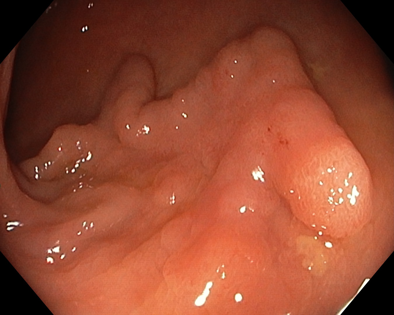
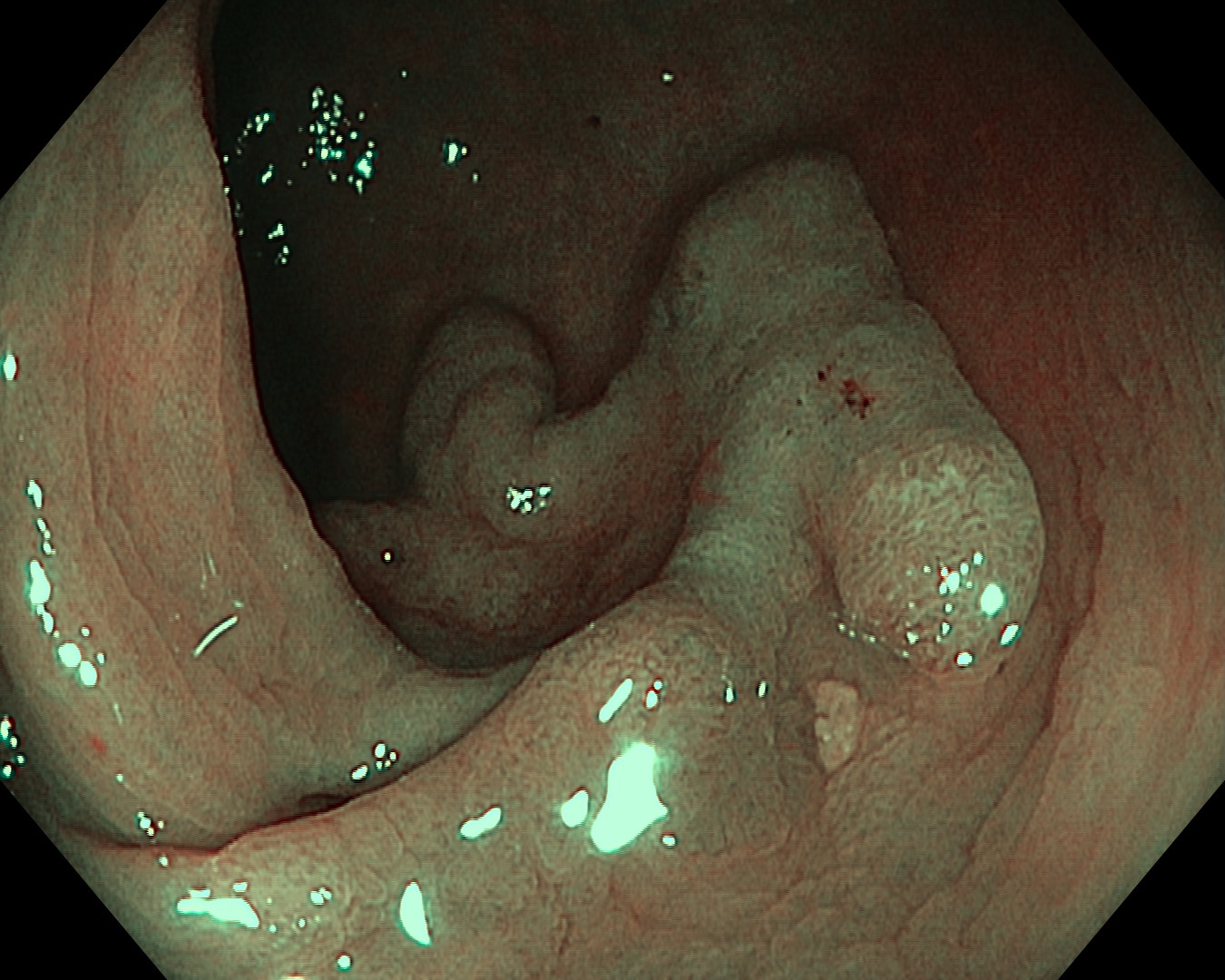
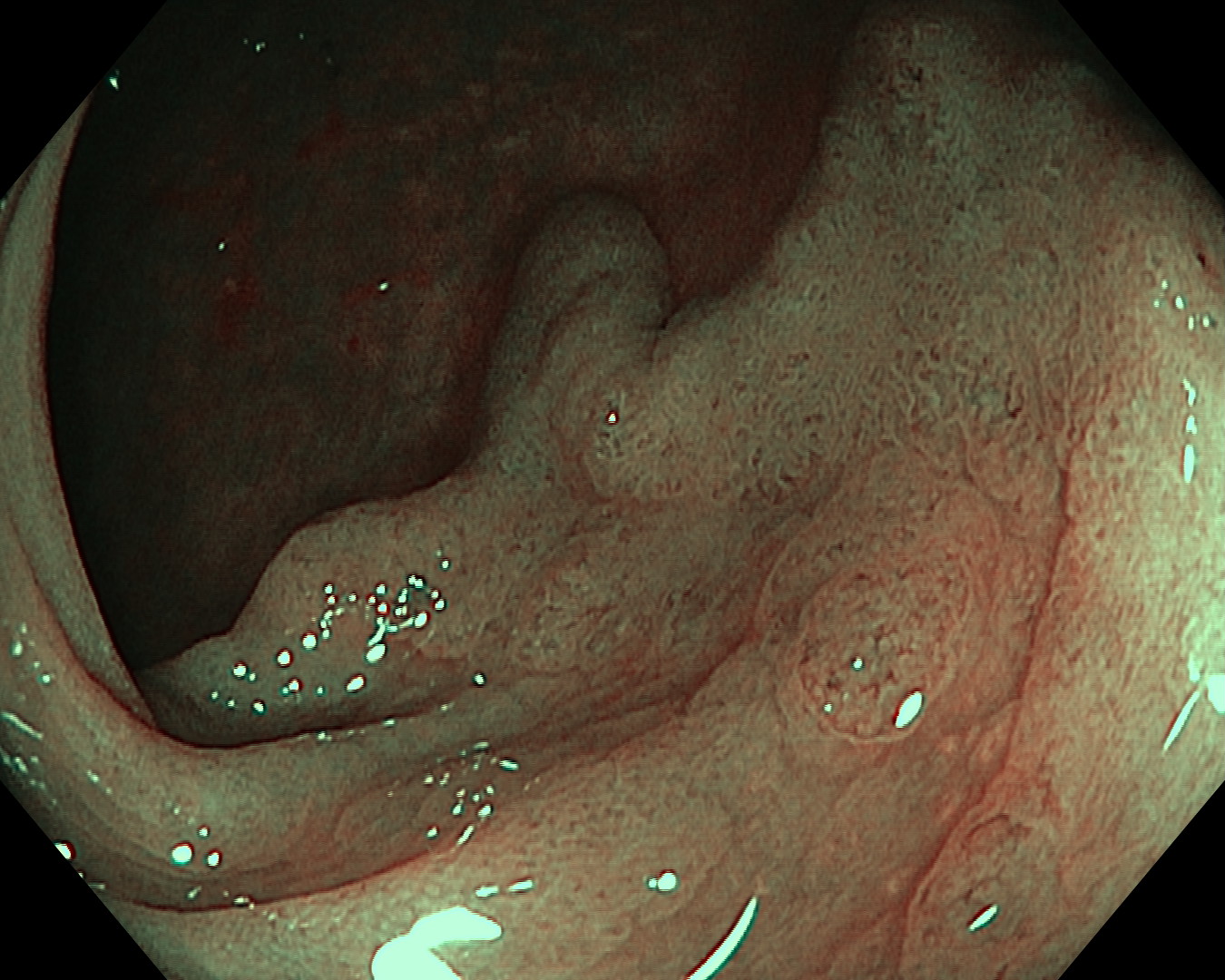
1. WLI Observation
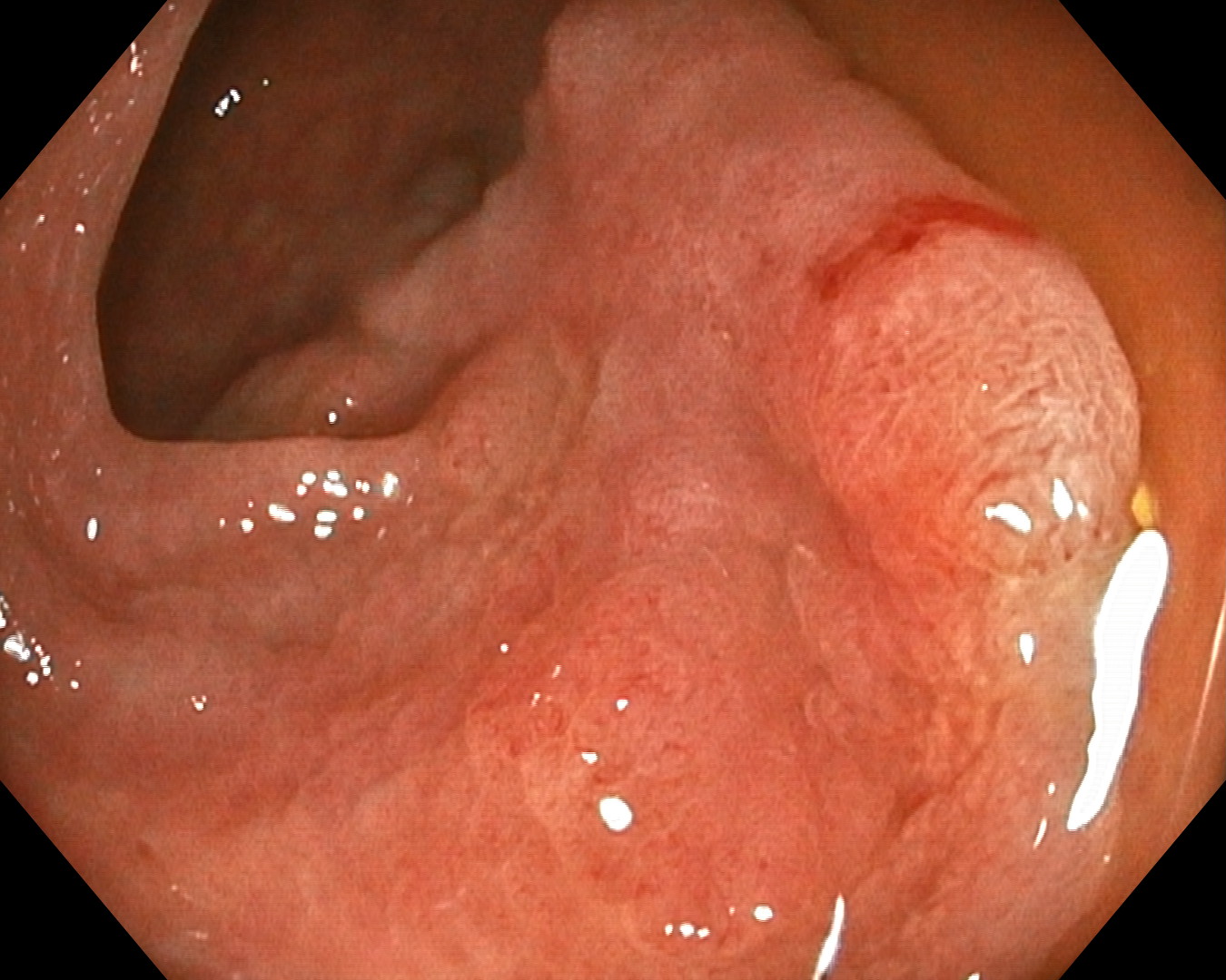
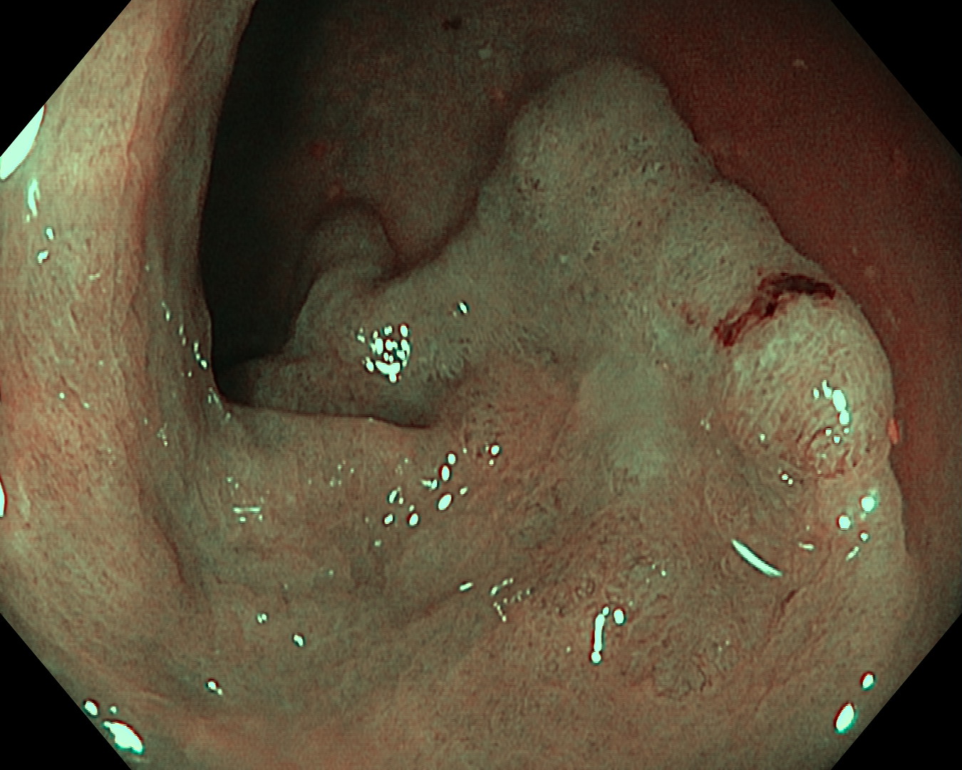
2. TXI Observation
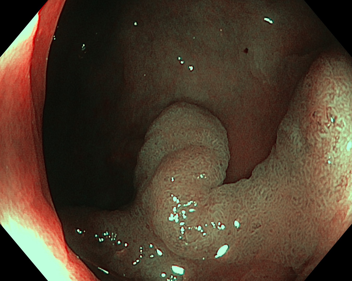
3. NBI Observation
4. NBI Observation
5. NBI Observation
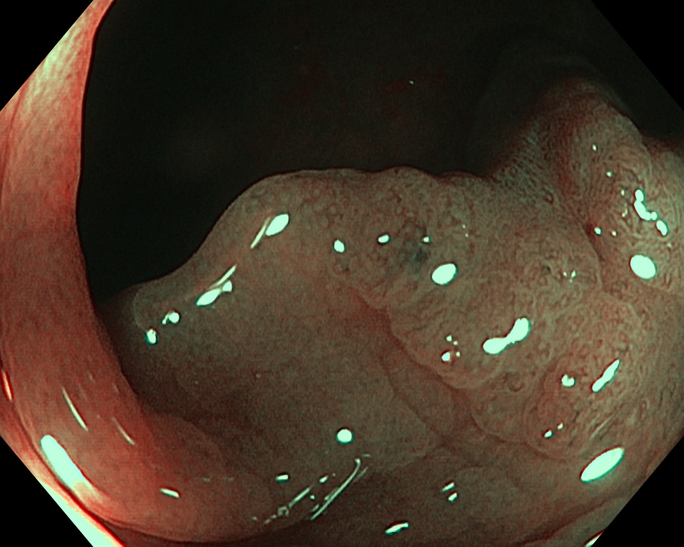
6. NBI Observation
7. NBI Observation
8. NBI Observation
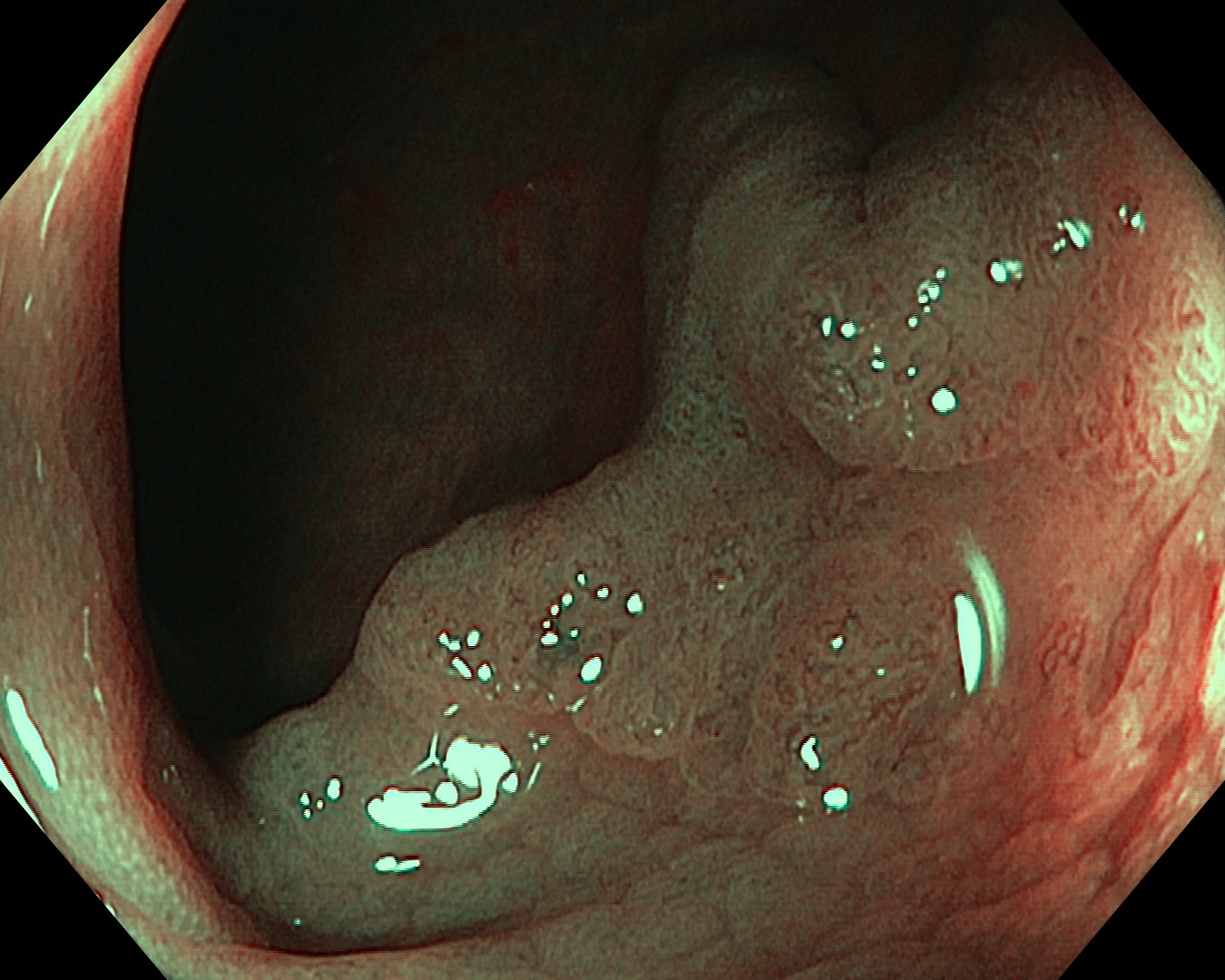
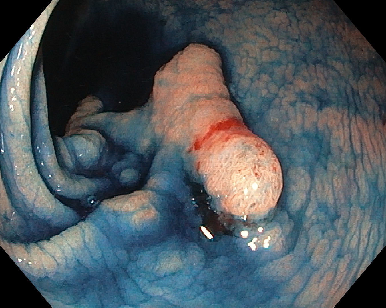
9. Indigocarmine Chromoendoscopy
Case Video
Overall Comment
The lesion was a flat elevated non-granular lateral spreading tumor measuring about 40mm at around the splenic flexure. Part of the lesion had indistinct border. Despite the inability to perform near focus examination due to the use of pediatric colonoscope, closed-up view under NBI was able to provide certain magnification of this lesion and it helped in border delineation and recognition of the superficial vessel type under JNET classification. Conventional chromoendoscopy using indigocarmine dye further aided the border delineation. This lesion will be subjected for endoscopic submucosal dissection.
* Specifications, design and accessories are subject to change without any notice or obligation on the part of the manufacturer
Dr. Shiaw-Hooi Ho Case 27: Intramucosal Carcinoma (High-grade Dysplasia)
Prof. Yoji Takeuchi
- Keyword
- Content Type