Colorectal Case 24

Dr. Shiaw-Hooi Ho
Associate Professor of Medicine at the Department of Medicine,
Universiti Malaya, Malaysia
Scope: CF-HQ190L
Organ: Rectum
Patient information: 52 years old, Male
Medical history: Intermittent painless per rectal bleeding past 2 years; known ocular myasthenia gravis
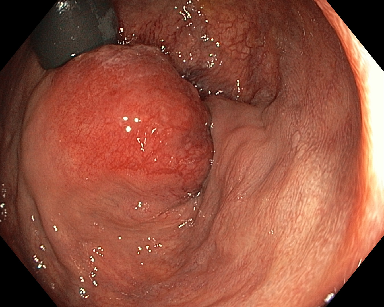
1. WLI Observation
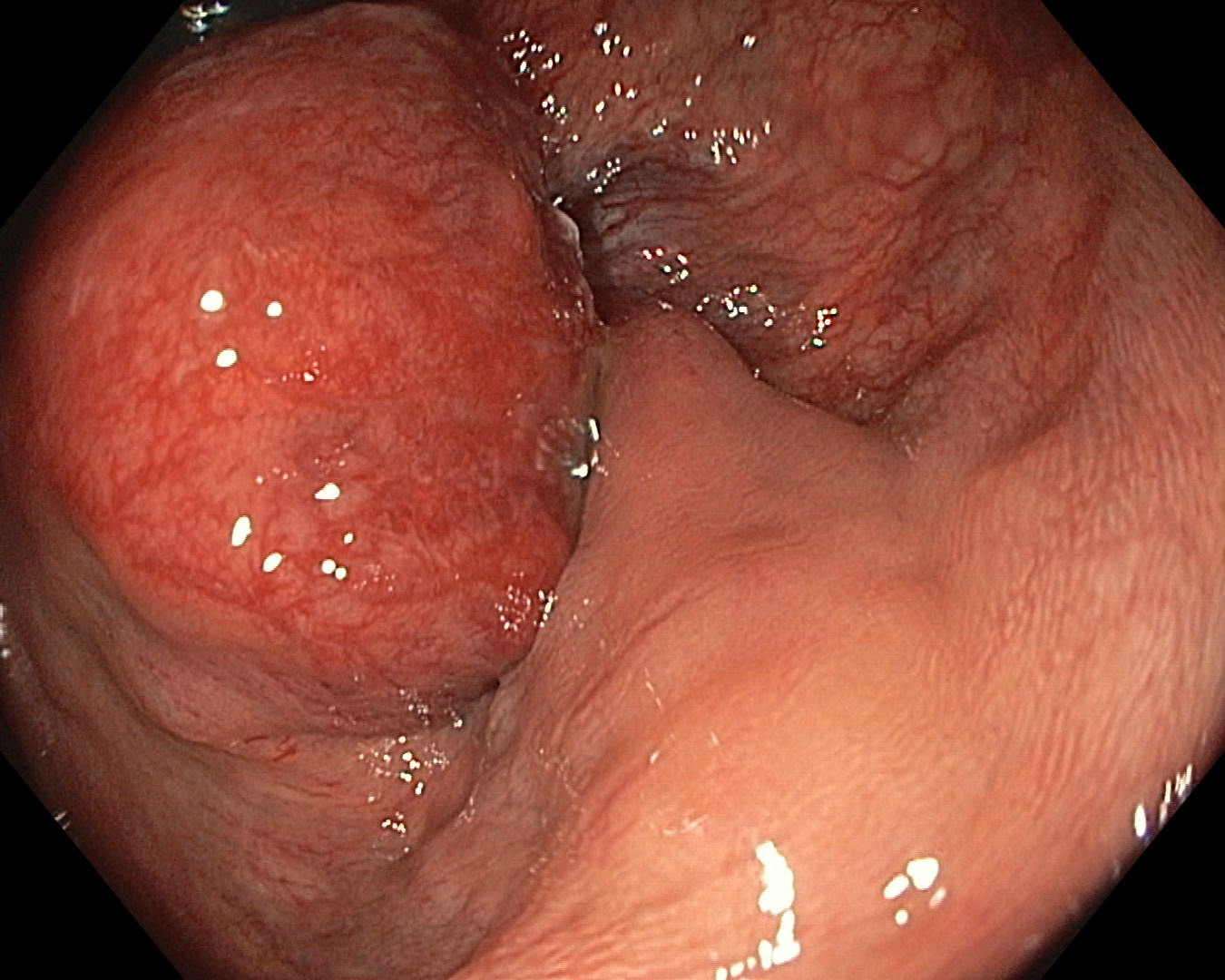
2. WLI Observation
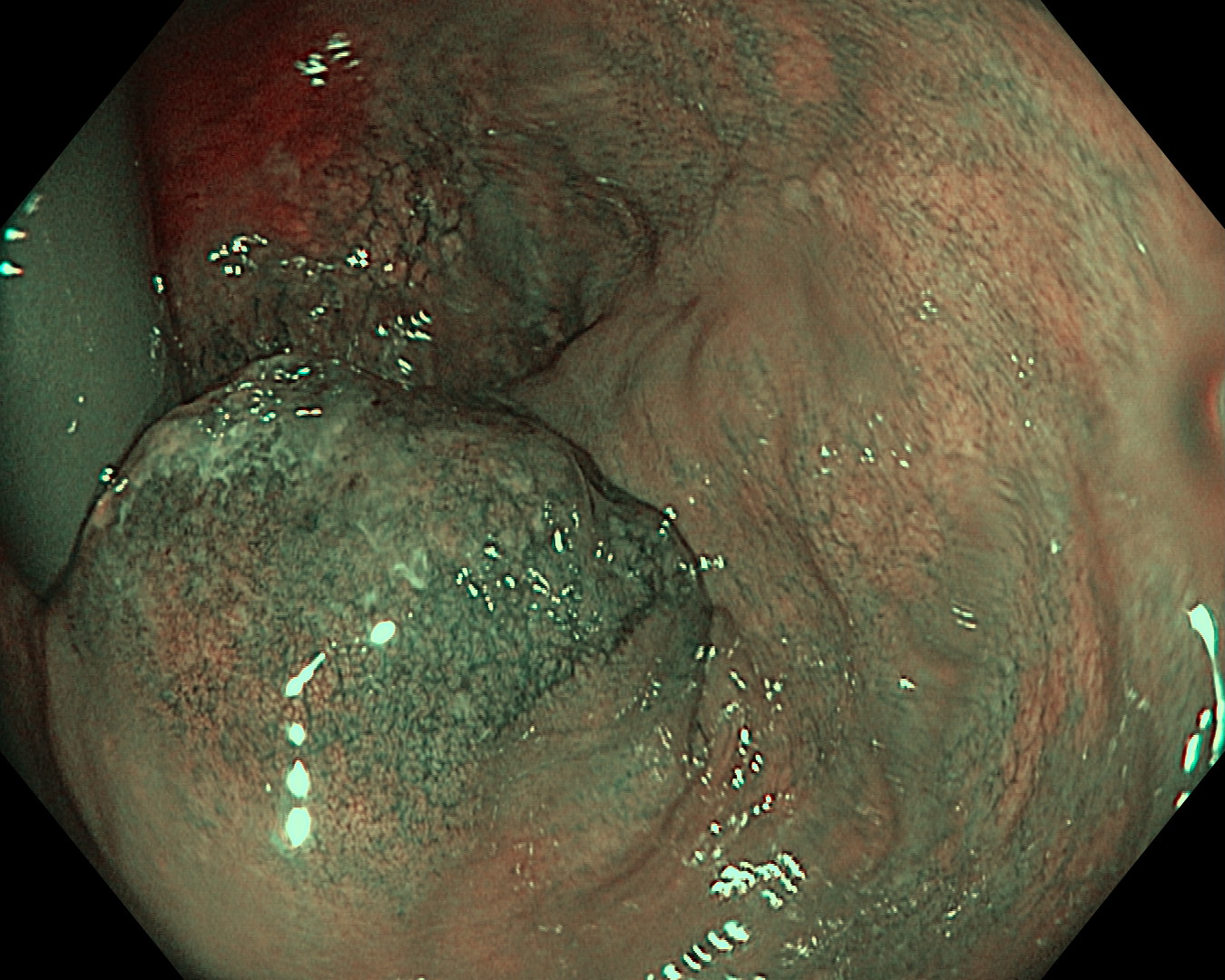
3. NBI Observation
Overall Comment
This patient was referred to colorectal surgical team for endoanal ultrasound examination and further management. He underwent trans-anal resection and further hemorrhoidectomy. The histology revealed hemorrhoid with stromal hemorrhage. There was no other abnormal tissue seen. This case illustrated an atypical presentation of an internal hemorrhoid with stromal hemorrhage mimicking a submucosal tumor. Other differential could be thrombosed hemorrhoid but there was no complaint of pain in this patient.
* Specifications, design and accessories are subject to change without any notice or obligation on the part of the manufacturer
Prof. Han-Mo Chiu Case 25: Crohn’s Ileo-colitis
Dr. Shiaw-Hooi Ho
- Keyword
- Content Type