Gastric Case 4

Dr. D Nageshwar Reddy
AIG Hospitals, Hyderabad, India

Dr. Hardik Rughwani
AIG Hospitals, Hyderabad, India
Scope: GIF-EZ1500
Case: Early gastric cancer
Organ: Stomach
Patient information: F, 76
Medical history: History of chronic dyspepsia and recent onset pain abdomen and weight loss
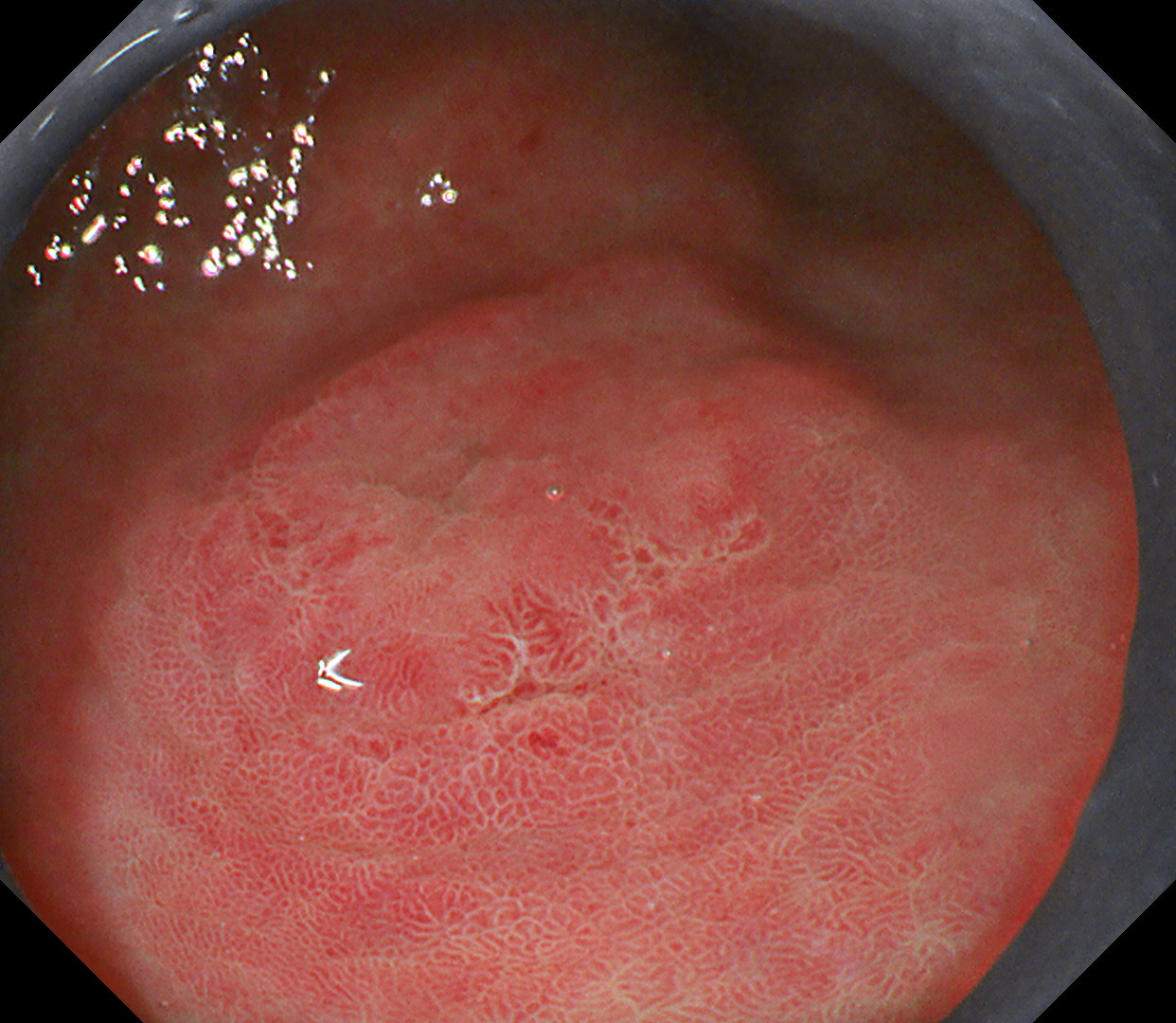
1. WLI
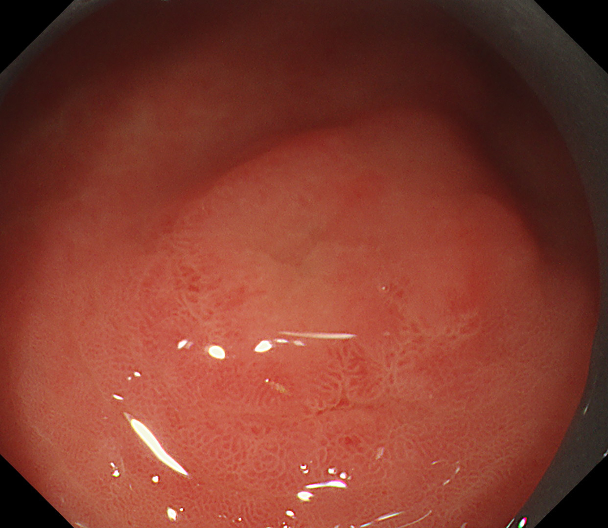
Single nodular lesion seen at antrum with elevated edges and indistinct margins. The demarcation line is not clearly visible in WLI.
2. TXI
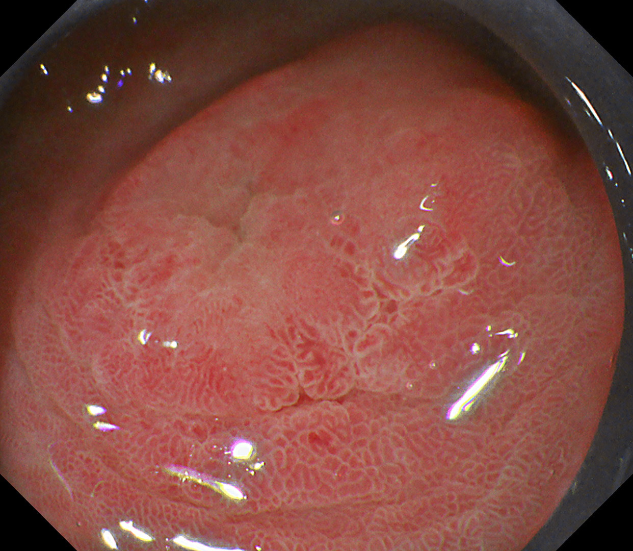
Upon TXI assessment, the border of the lesion is clearly seen with irregular shape and color, suspicious of neoplastic etiology. The demarcation line is clearly visible.
3. NBI
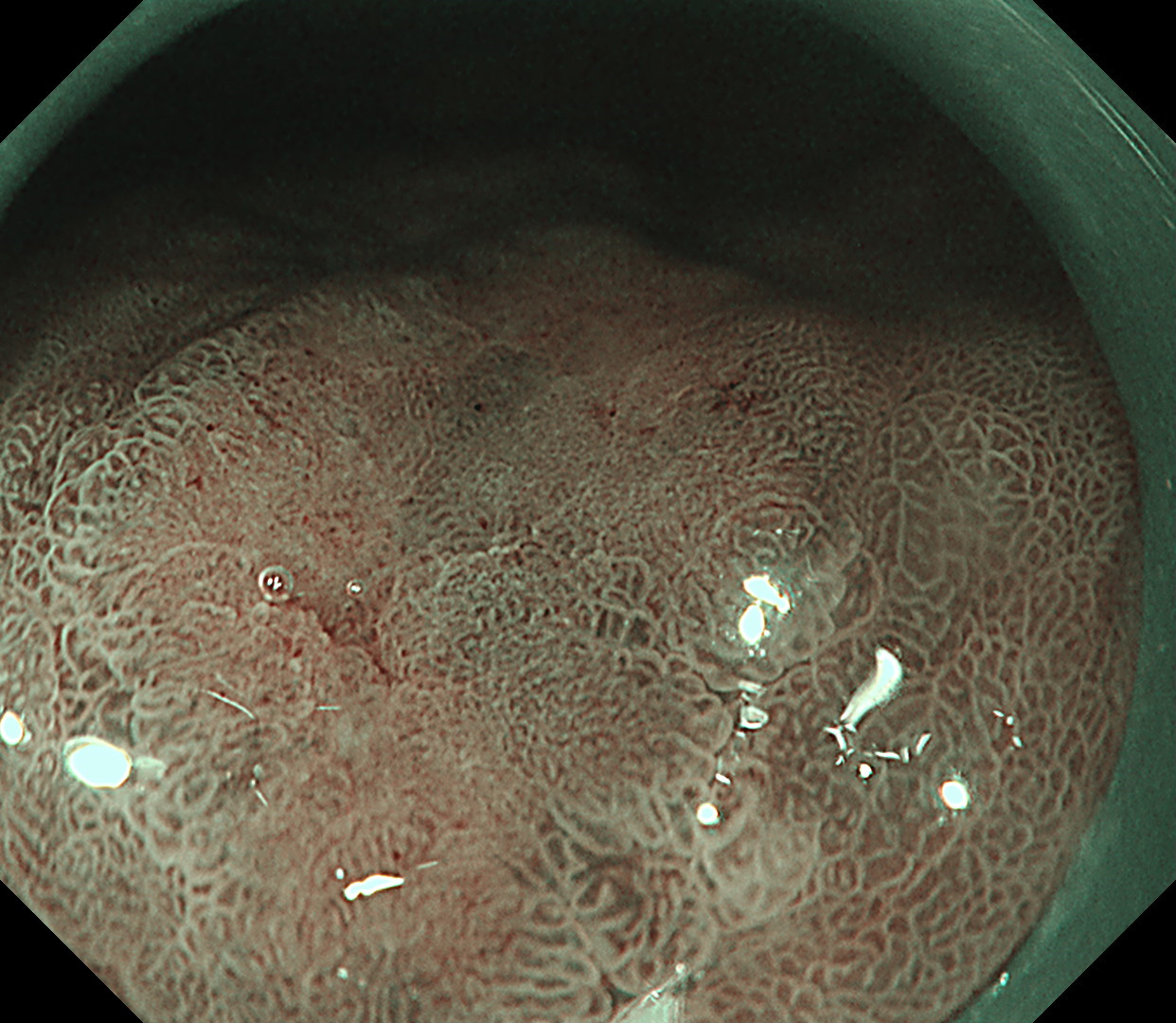
NBI with magnification demonstrated clear demarcation line (DL), near the lesion, with irregular micro surface (MS) and microvascular pattern (MV) s/o Early gastric cancer. Light blue crest (LBC) sign on epithelial surface is s/o Intestinal Metaplasia.
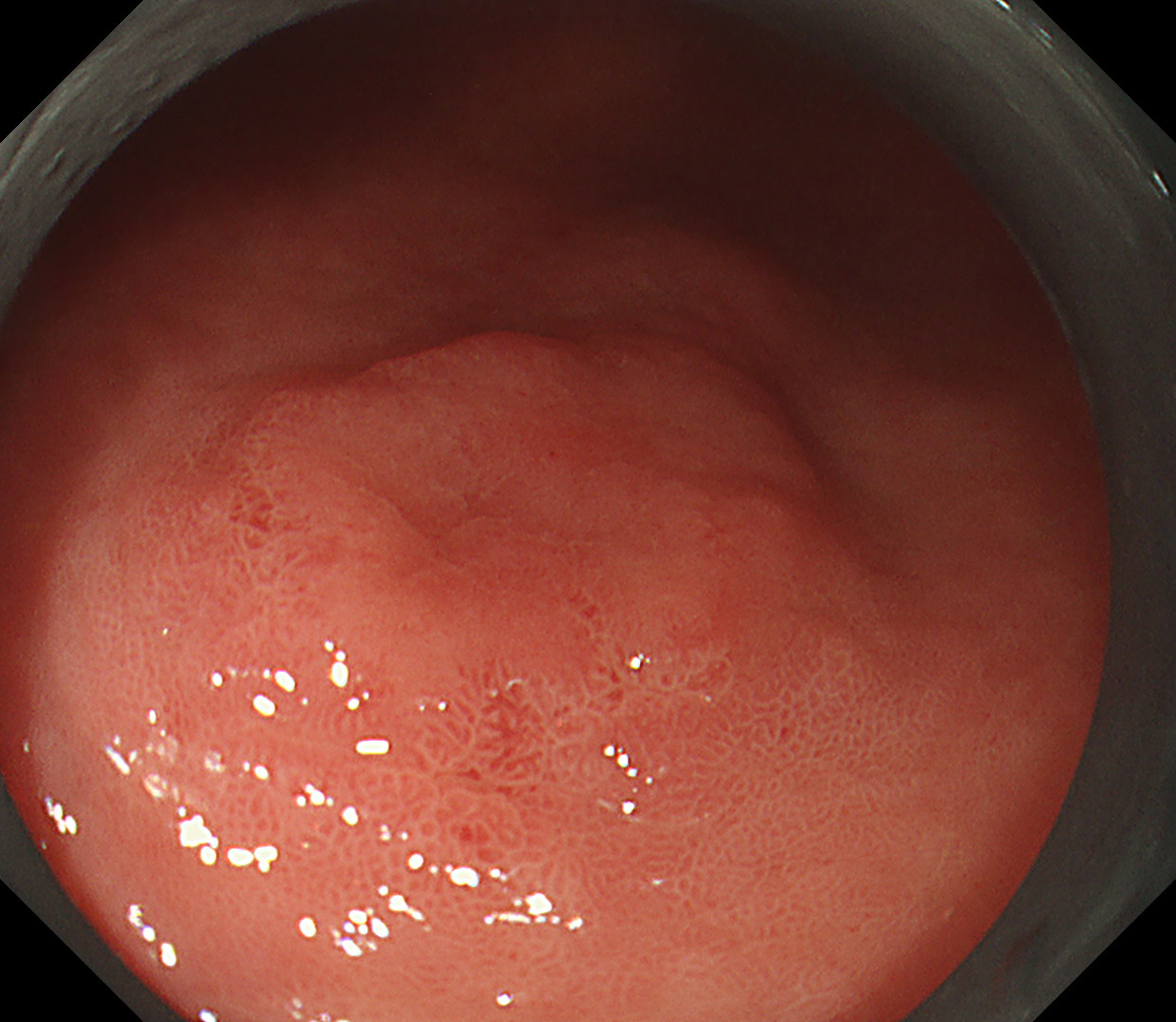
4. WLI
Single nodular lesion seen at antrum with elevated edges and indistinct margins. The demarcation line is not clearly visible in WLI.
5. TXI
Upon TXI assessment, the border of the lesion is clearly seen with irregular shape and color, suspicious of neoplastic etiology. The demarcation line is clearly visible
6. NBI
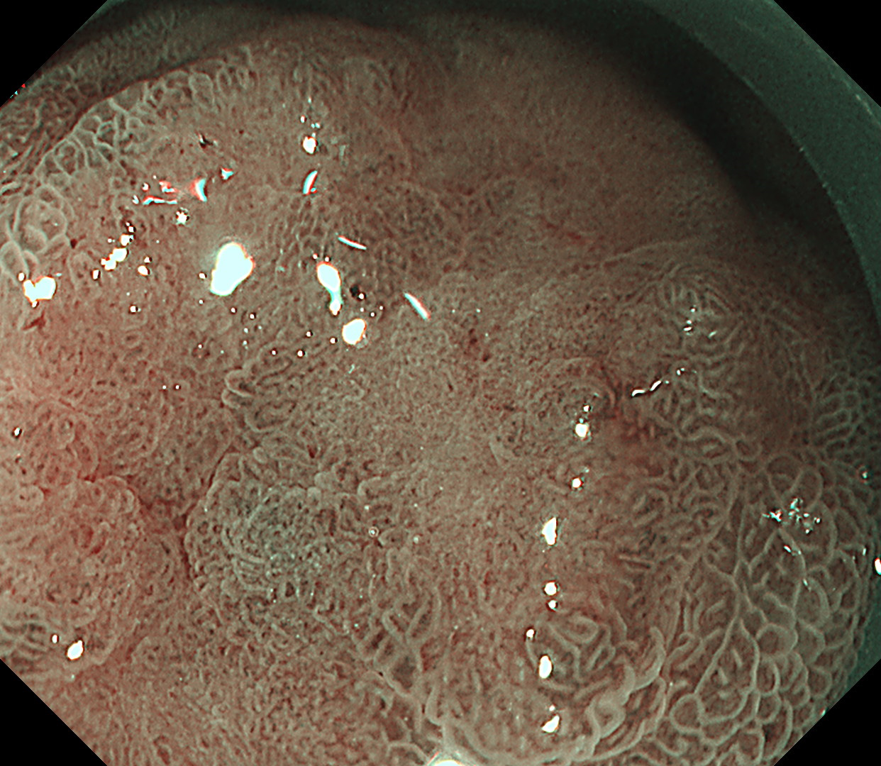
NBI with magnification demonstrated clear demarcation line (DL), near the lesion, with irregular micro surface (MS) and microvascular pattern (MV) s/o Early gastric cancer. Light blue crest (LBC) sign on epithelial surface is s/o Intestinal Metaplasia.
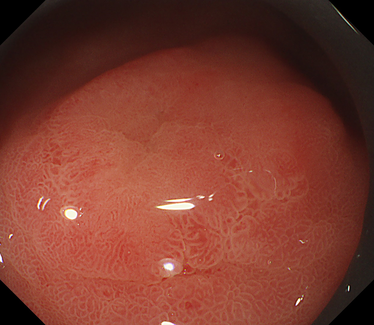
7. TXI mode 1
Upon TXI assessment, the border of the lesion is clearly seen with irregular shape and color, suspicious of neoplastic etiology. The demarcation line is clearly visible.
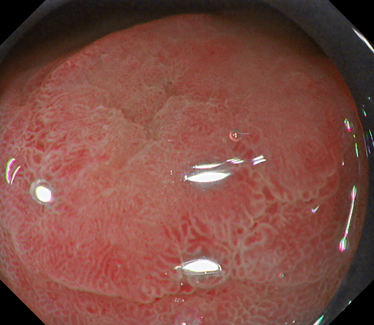
8. TXI Mode 2
TXI mode 2 of the same WLI shows enhancement of brightness and texture compared to WLI. The naturalness of the mode 2 image is better than the mode 1 image.
Overall Comment
TXI can enhance brightness selectively in dark areas of endoscopic images and can enhance subtle tissue differences such as slight morphological and/or color changes over conventional enhancement methods including structure enhancement and color enhancement. Comparing TXI with conventional white light imaging (WLI) for real-world image processing, TXI can achieve significant image enhancement with characteristics of WLI appearance while preventing over-enhancement observed in other methods. TXI has two modes, mode1 and mode 2. Enhancement of brightness and texture is similar in both modes. The enhancement of color contrast of mode1 is superior to that of mode 2 in accordance with the color enhancement of the mode1 algorithm; however, the naturalness in mode 2 is better than that of mode 1. Overall, TXI provides a balance of improving image features important for the physician searching for abnormalities (selective increase in brightness, greater color separation, and texture enhancement) while minimizing gross changes that may negatively impact familiarity (naturalness).
* Specifications, design and accessories are subject to change without any notice or obligation on the part of the manufacturer.
Dr. D Nageshwar Reddy
Dr. Hardik Rughwani Case 5: Low grade dysplasia (Duodenum)
- Content Type