Gastric Case 3
Dr. D Nageshwar Reddy
AIG Hospitals, Hyderabad, India
Dr. Hardik Rughwani
AIG Hospitals, Hyderabad, India
Scope: GIF-EZ1500
Case: Multiple gastric polyps (Benign)
Organ: Stomach
Patient information: F, 50
Medical history: History of chronic iron deficiency anemia and dyspepsia
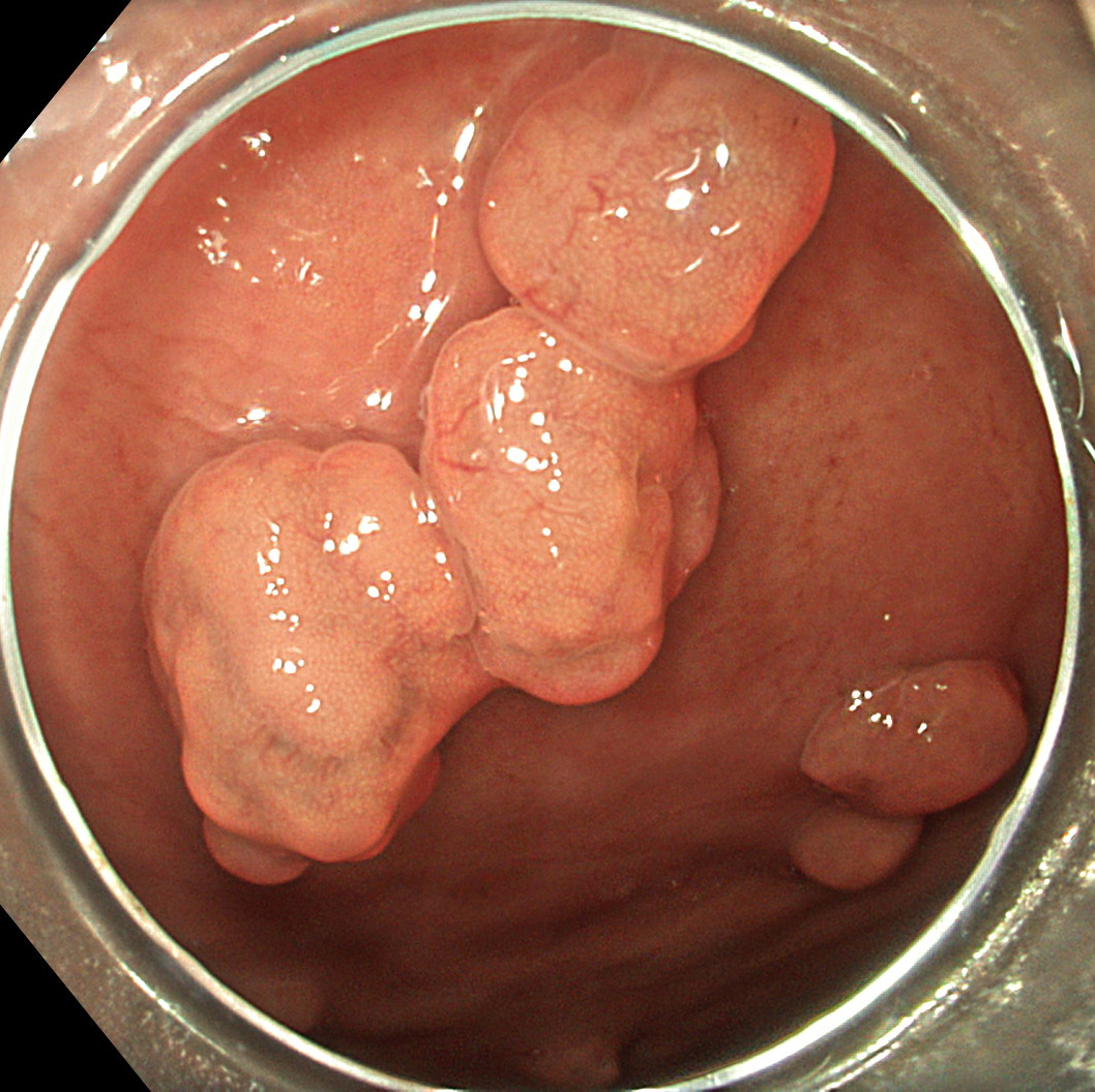
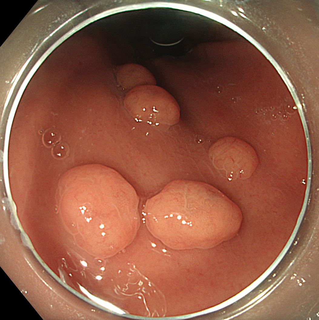
1. WLI
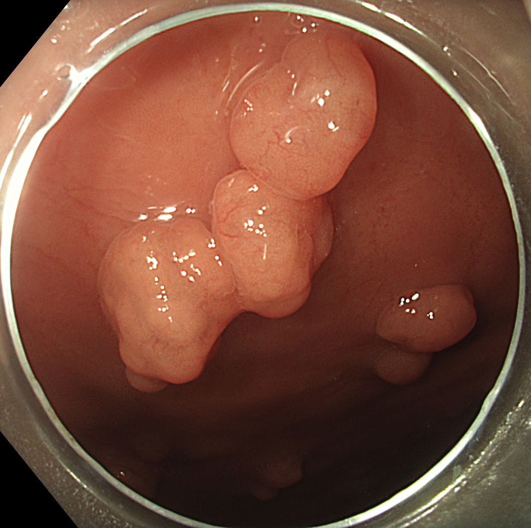
Multiple sessile polyps of <1cm sizes noted in the body of the stomach
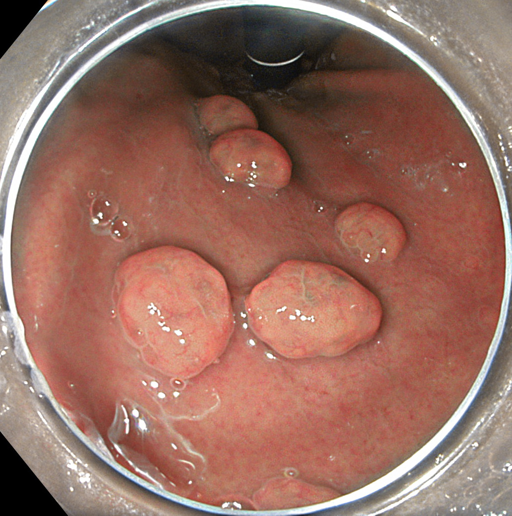
2. TXI mode 1
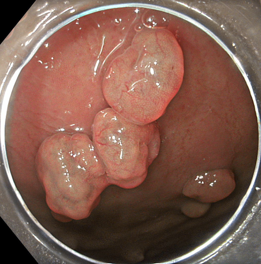
TXI mode 1 image of the same WLI shows better color and structural image enhancement compared to WLI. There is increased brightness in the darker areas of the WLI.
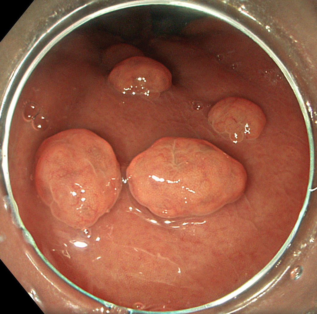
3. TXI mode 2
TXI mode 2 of the same WLI shows enhancement of brightness and texture compared to WLI. The naturalness of the mode 2 image is better than the mode 1 image.
4. WLI
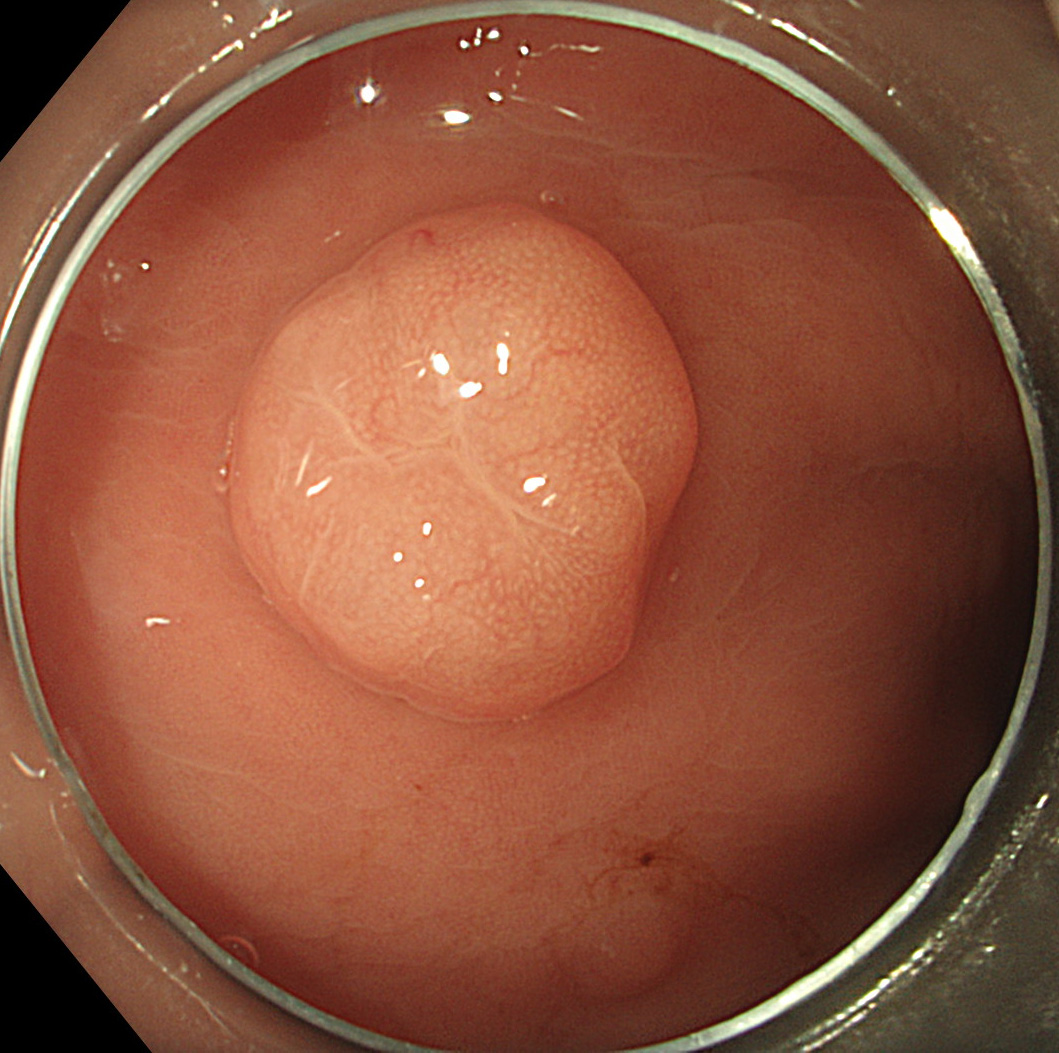
Single sessile polyp of >1cm size noted in the body of the stomach
5. TXI mode 1
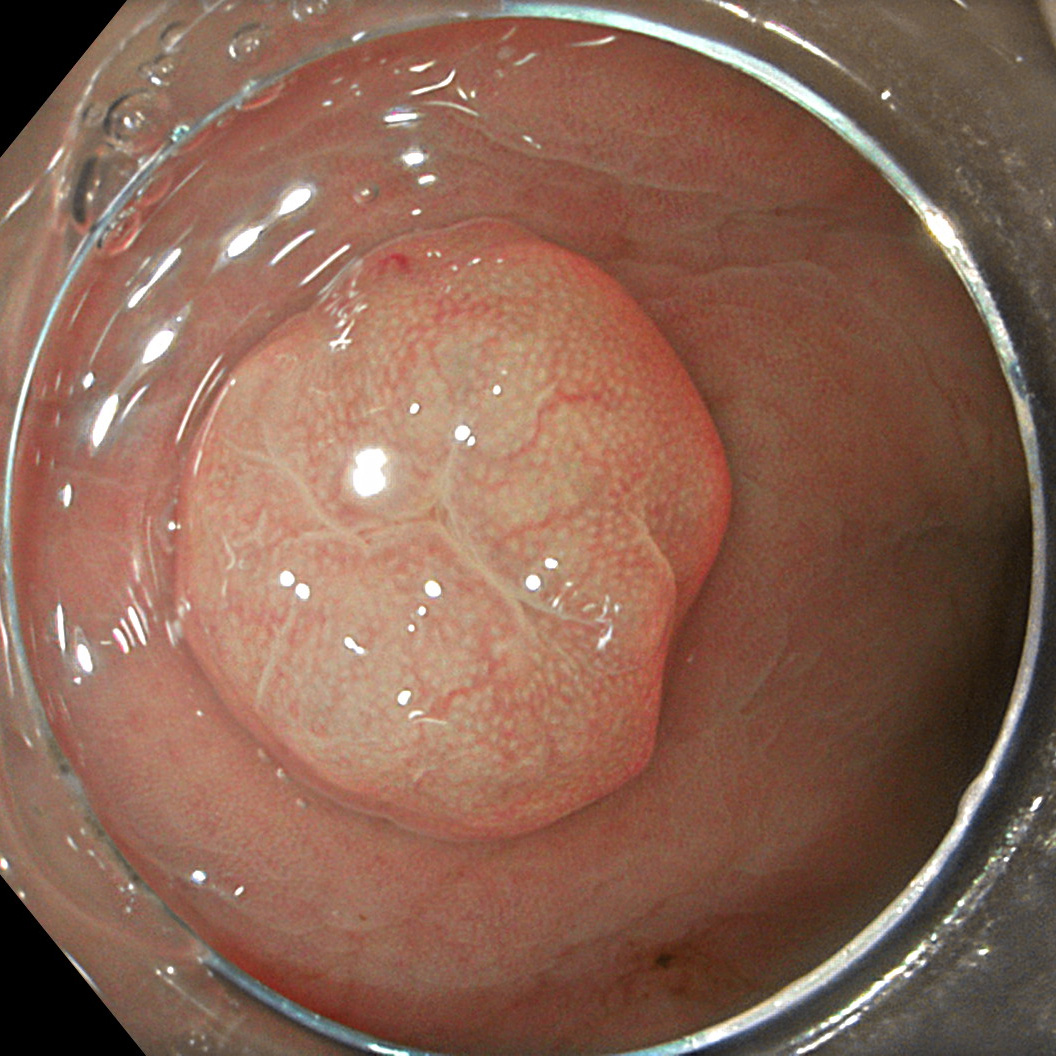
TXI mode 1 image of the same WLI shows better color and structural image enhancement compared to WLI. There is increased brightness in the darker areas of the WLI.
6. TXI mode 2
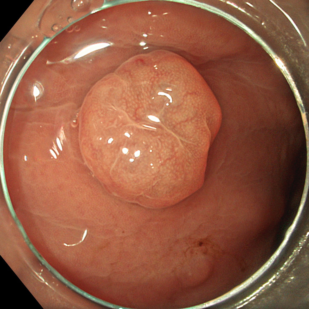
TXI mode 2 of the same WLI shows enhancement of brightness and texture compared to WLI. The naturalness of the mode 2 image is better than the mode 1 image.
7. WLI
Multiple sessile polyps of <1cm sizes noted in the body of the stomach
8. TXI mode 1
TXI mode 1 image of the same WLI shows better color and structural image enhancement compared to WLI. There is increased brightness in the darker areas of the WLI.
9. TXI mode 2
TXI mode 2 of the same WLI shows enhancement of brightness and texture compared to WLI. The naturalness of the mode 2 image is better than the mode 1 image.
Overall Comment
TXI can enhance brightness selectively in dark areas of endoscopic images and can enhance subtle tissue differences such as slight morphological and/or color changes over conventional enhancement methods including structure enhancement and color enhancement. Comparing TXI with conventional white light imaging (WLI) for real-world image processing, TXI can achieve significant image enhancement with characteristics of WLI appearance while preventing over-enhancement observed in other methods. TXI has two modes, mode1 and mode 2. Enhancement of brightness and texture is similar in both modes. The enhancement of color contrast of mode1 is superior to that of mode 2 in accordance with the color enhancement of the mode1 algorithm; however, the naturalness in mode 2 is better than that of mode 1. Overall, TXI provides a balance of improving image features important for the physician searching for abnormalities (selective increase in brightness, greater color separation, and texture enhancement) while minimizing gross changes that may negatively impact familiarity (naturalness).
* Specifications, design and accessories are subject to change without any notice or obligation on the part of the manufacturer.
Prof. Yip Hon Chi Case 4: Early gastric cancer
Dr. D Nageshwar Reddy
Dr. Hardik Rughwani
- Content Type


