Prof. Jean de la Rosette, MD. Academic Medical Centre, Netherlands
Multiple papillary tumors age 74, male
White Light
NBI
White Light
NBI
White Light
NBI
White Light
NBI
White Light
NBI
Comments
Multiple papillary lesions, especially on the bladder trigone, posterior and anterior wall were visible with WLI.
After NBI-enhancement, additional multiple papillary fields were visualized. Histology showed pTa, Low grade (G1).
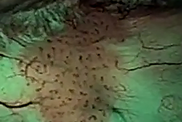
Multiple papillary tumors age 88, male
White Light
NBI
Comments
Multiple papillary lesions, especially on the left bladder wall and behind the right ostium,
clearly visible after NBI-enhancement. Histology showed pTa, Low grade (G1).
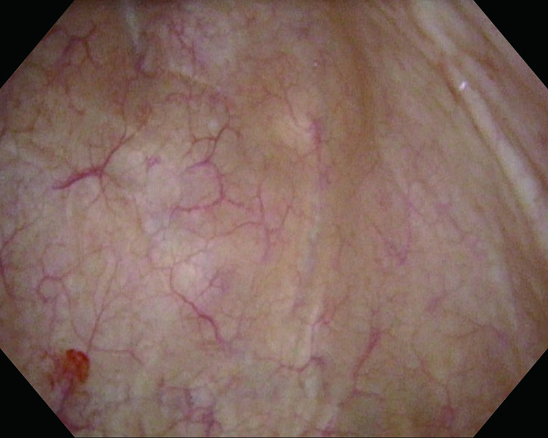
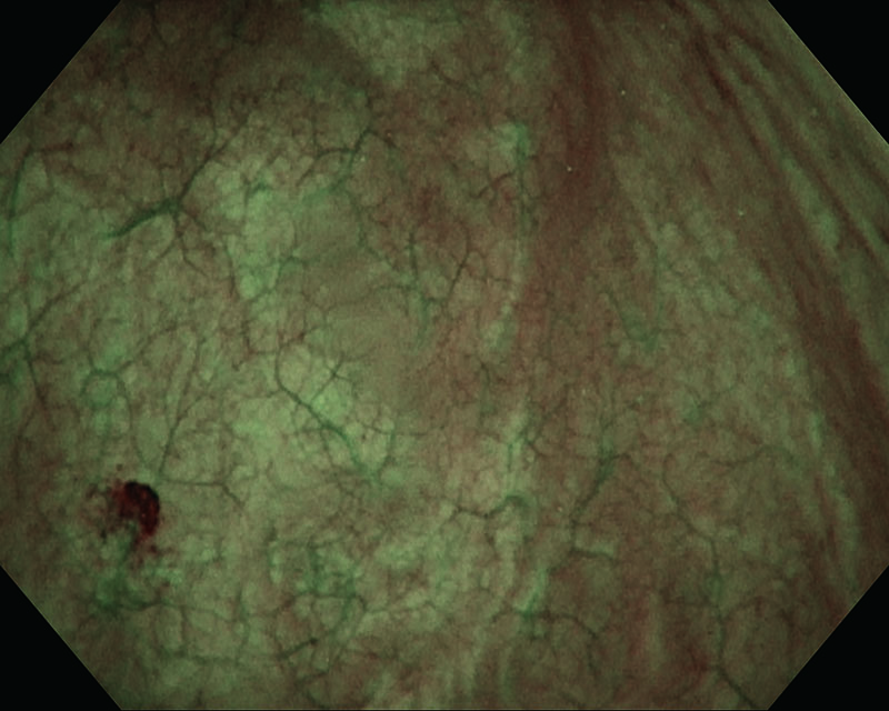
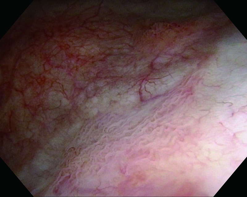
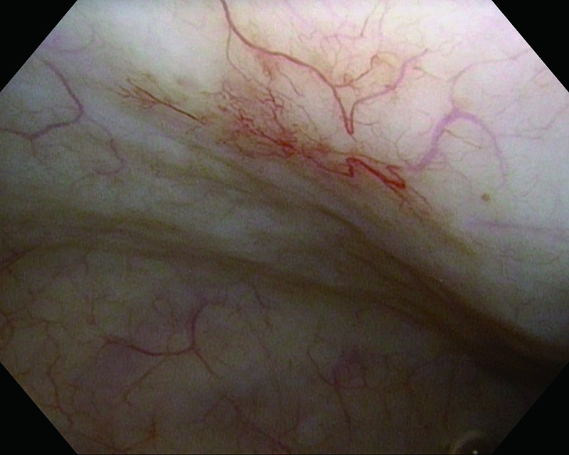
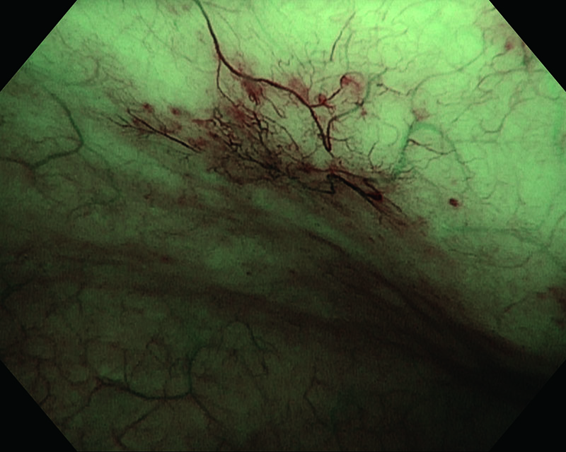
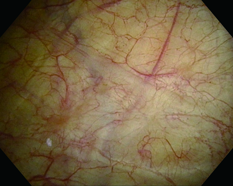
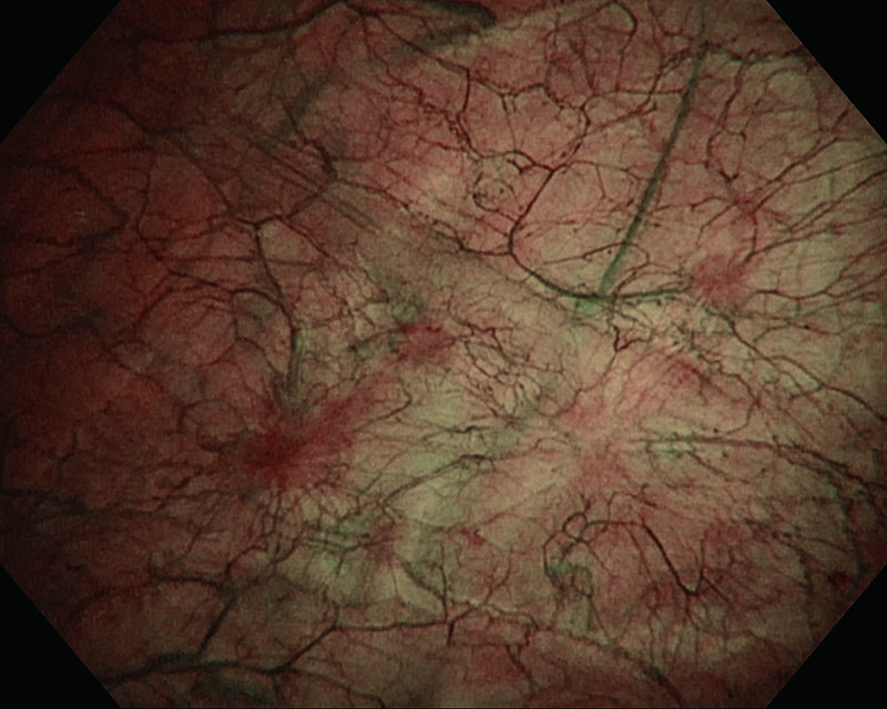
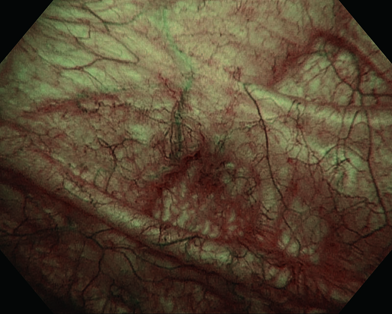
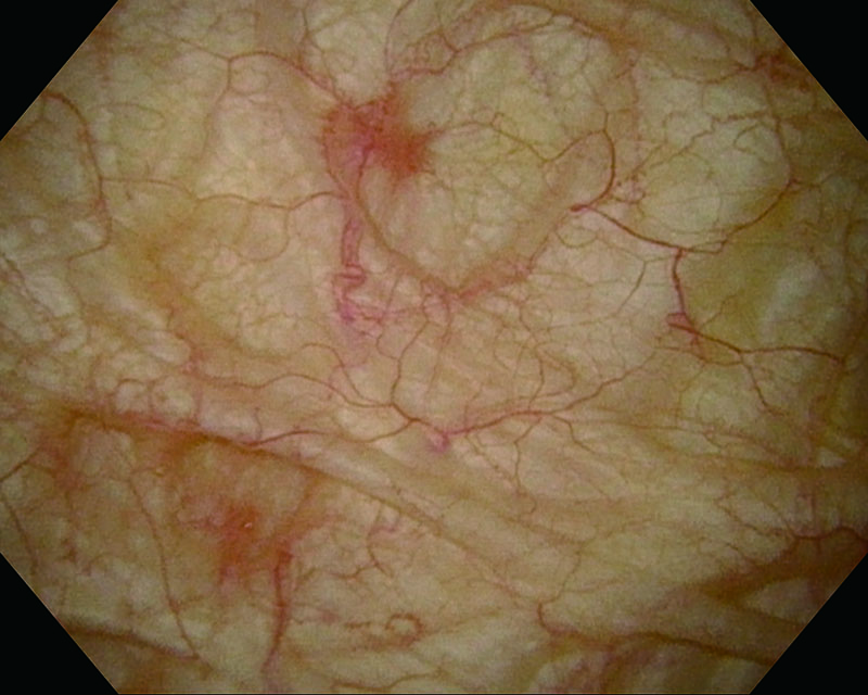
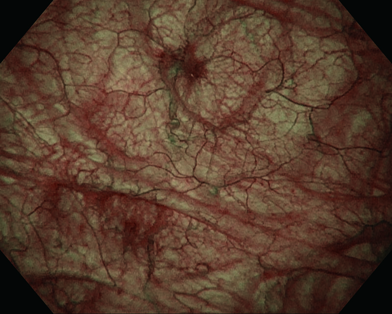
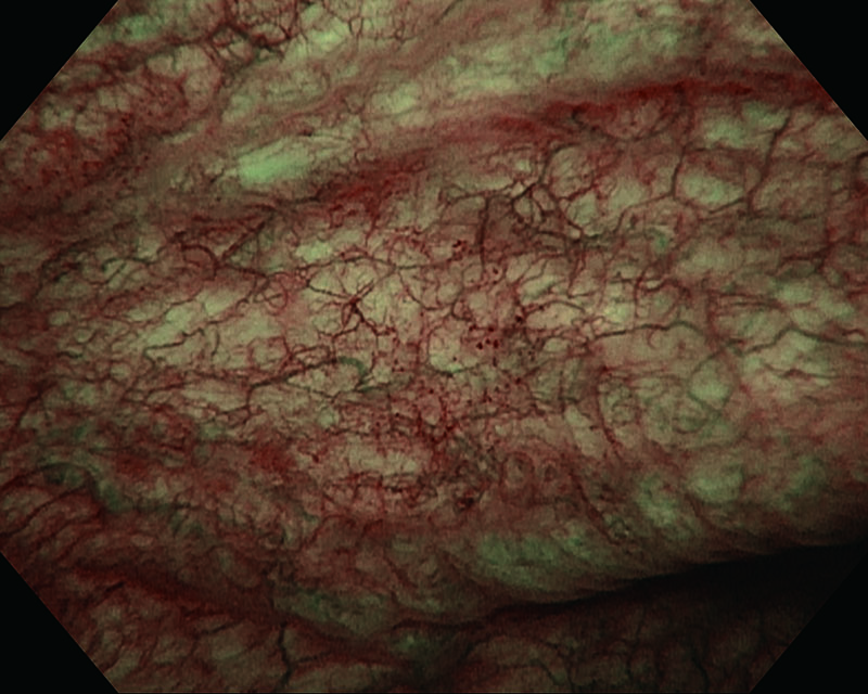
Congestive mucosa of the bladder's trigone age 28, female
White Light

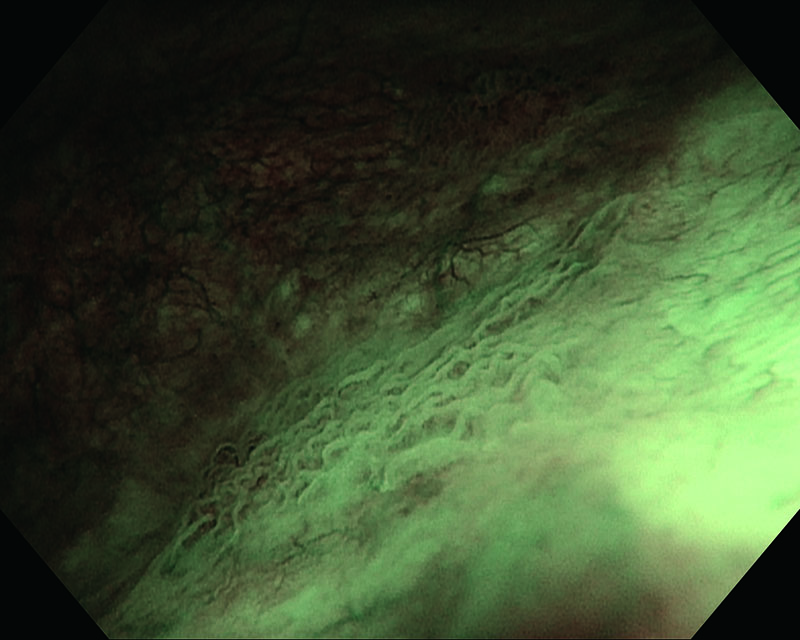
NBI
Comments
Congestive mucosa of the bladder trigone. NBI enhances the hypervascularized area. Histology showed pTa, Low grade (G2).
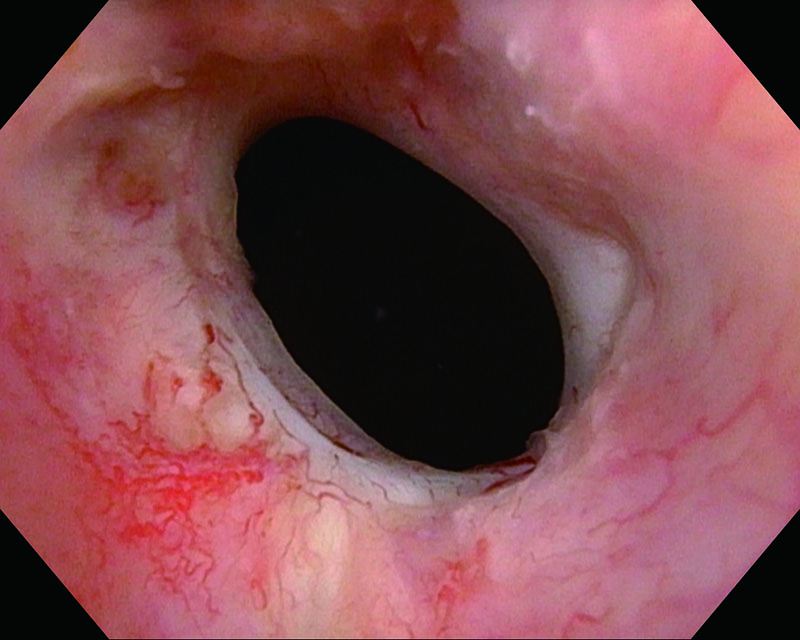
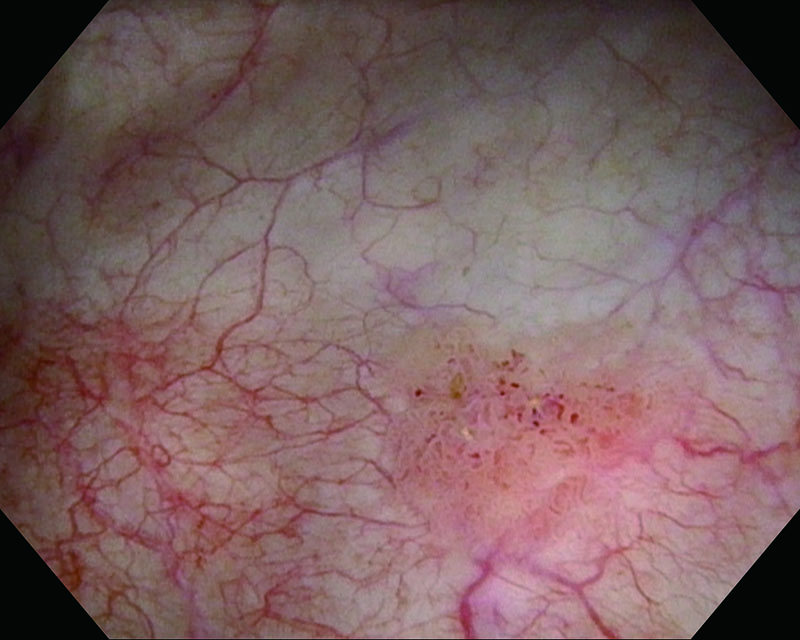
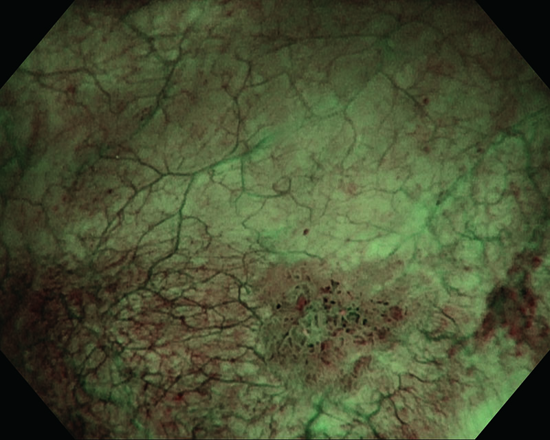
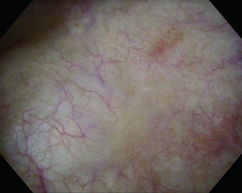
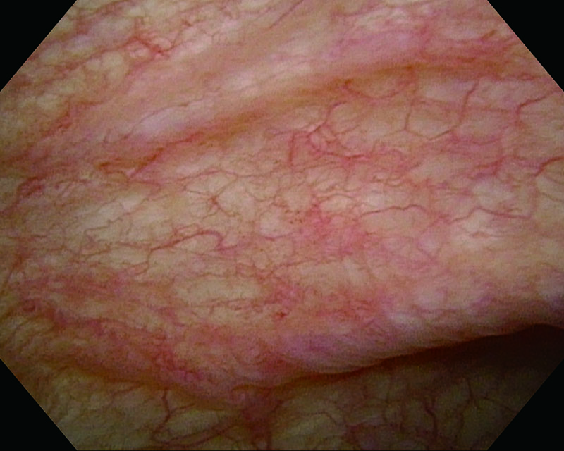
Flat Lesion age 69, male
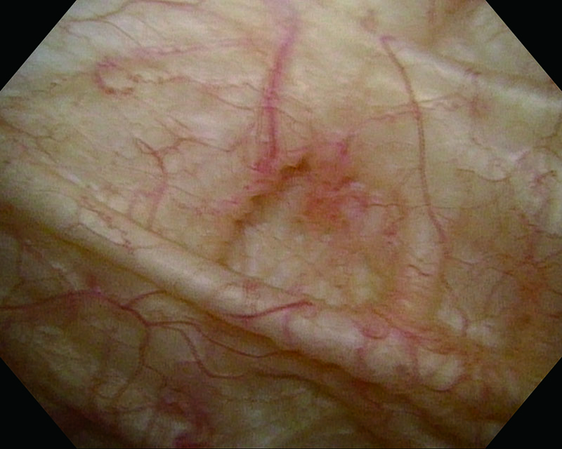
White Light
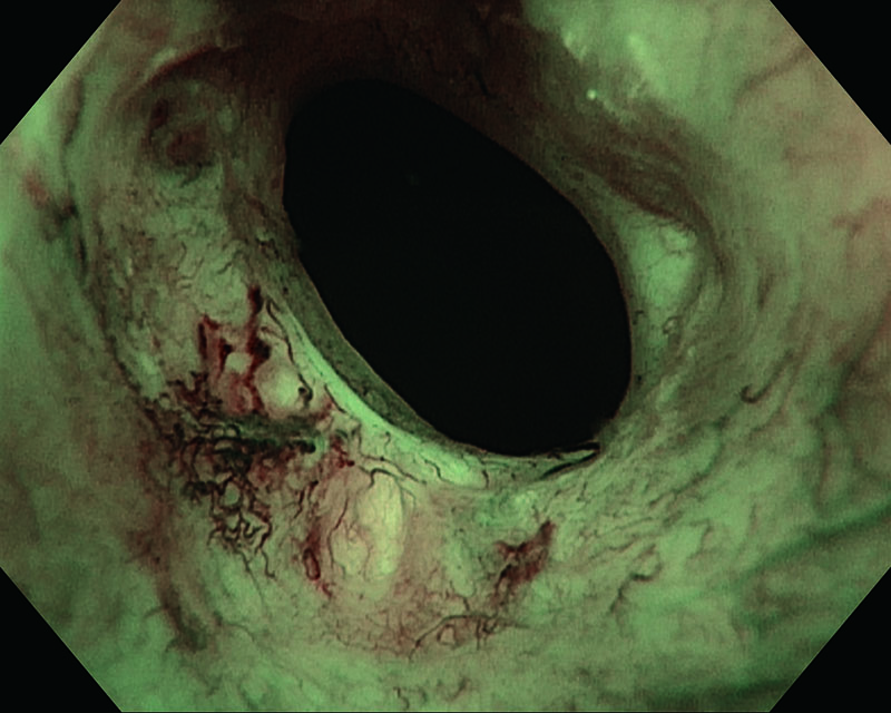
NBI
White Light
NBI
White Light
NBI
Comments
Suspicious superficial lesions adjacent to the right ostium, visible after NBI-enhancement.
Histology showed pTa, Low grade (G2).
Images and comments by Prof. Jean de la Rosette, MD.
Junichi Inokuchi, MD. Katsunori Tatsugami, MD. Prof. Seiji Naito, MD. Kyushu University, Japan
Angelo Naselli, MD. Prof. Paolo Puppo, MD. Oncological Urology, Istituto Clinico Humanitas Mater Domini, Castellanza, Varese, Italy
- Content Type