Cytology Atlas
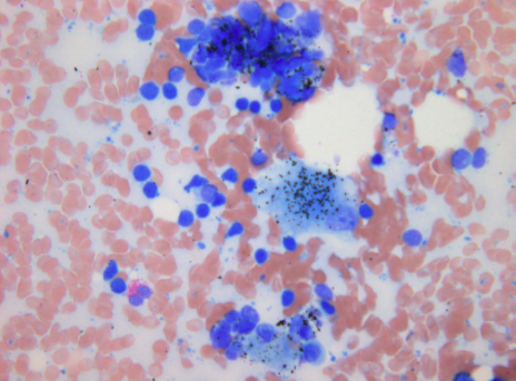
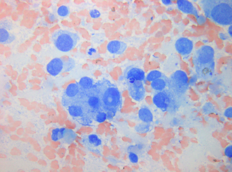
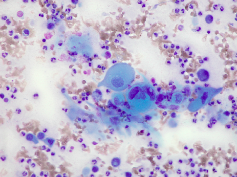
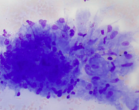
Representative EBUS‐TBNA from a lymph node with many benign‐appearing lymphocytes without any granulocytes and with anthracotic pigmentladen macrophages
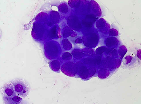
Representative lymph node sample (MGG)
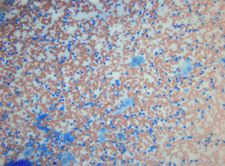
Representative lymph node sample (MGG)
Representative lymph node sample (MGG)
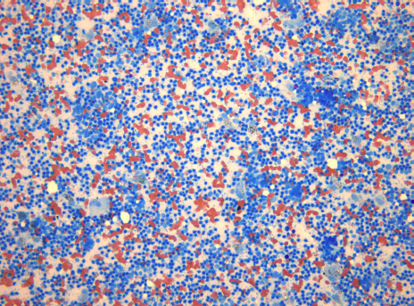
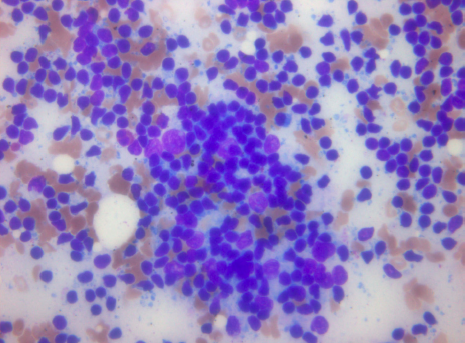
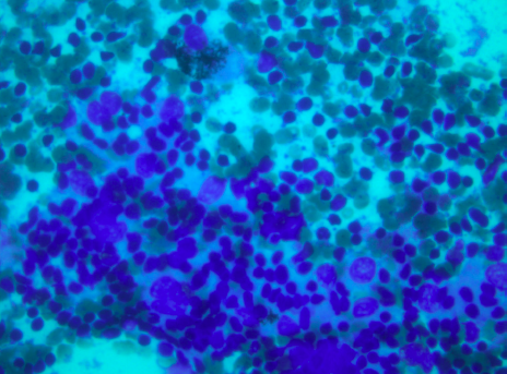
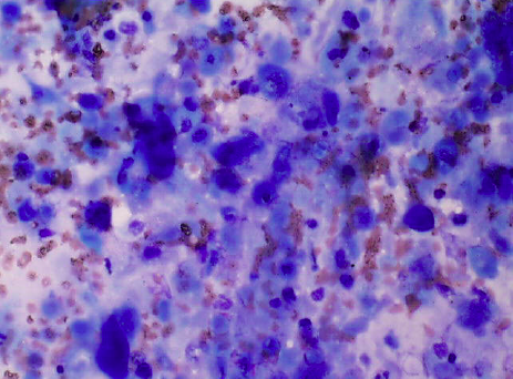
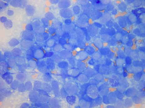
Reference Images - Lymphocytes
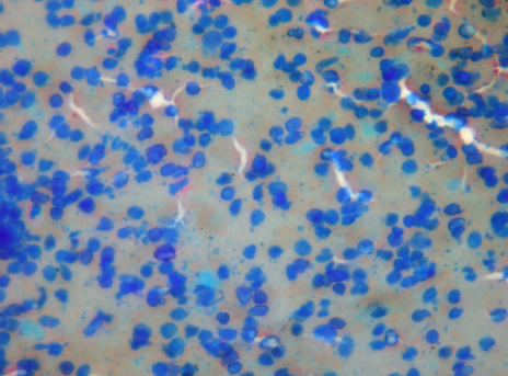
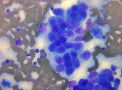
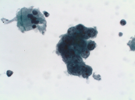
Clusters of admixed lymphocytes (MGG)
Representative lymph node sample (MGG)
Clusters of admixed lymphocytes (MGG)
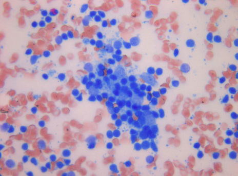
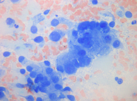
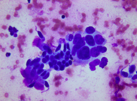
Cluster of admixed lymphocytes and pigmented macrophages (MGG)
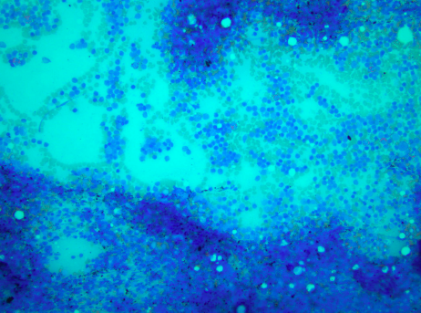
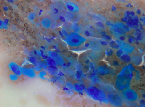
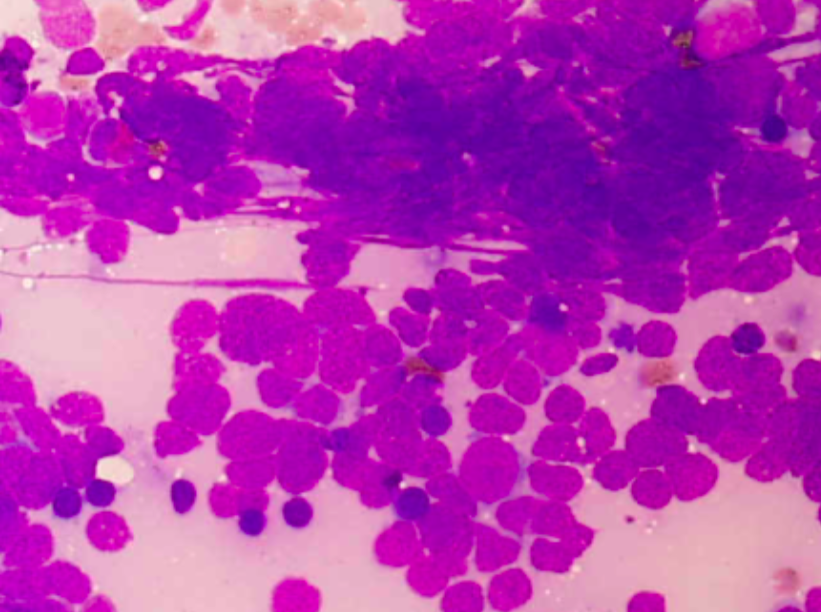
Reference Images – Lymphocytes and Anthracosis
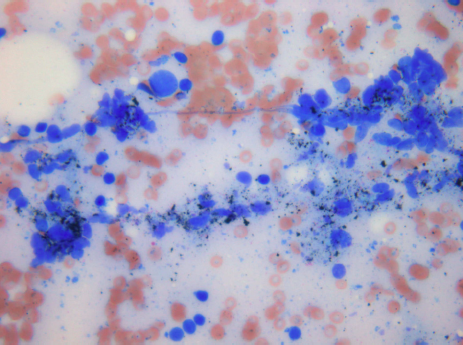
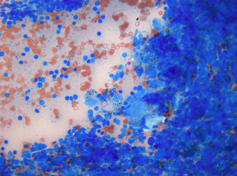
Lymph node sample with anthracosis (MGG)
Lymph node sample with anthracosis (MGG)
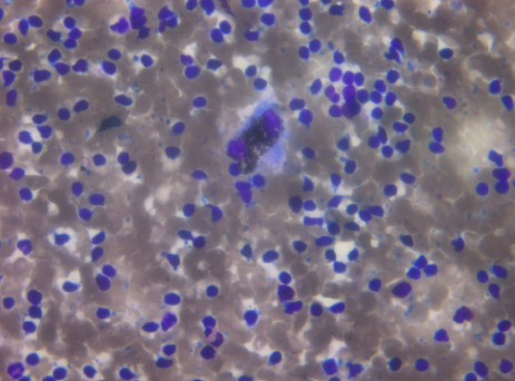
A representative EBUS‐TBNA from a lymph node with many benign‐appearing lympho‐cytes without any granulocytes and an anthracotic pigmentladen macrophage (MGG)
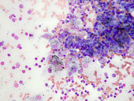
A lymph node sample with a large number of anthracotic pigment‐laden macrophages (MGG)
Reference Images – Adenocarcinoma
Adenocarcinoma (MGG)
Adenocarcinoma (MGG)
Adenocarcinoma (MGG)
A sheet of ciliated columnar, normal respiratoty epithe‐lium is visible in the lower left part of the image (MGG)
Adenocarcinoma cells are present in the center, with large, hyperchromatic nuclei, multiple angular nucleoli, and irregular nuclear envelopes (PAP), normal respiratory epithelium in the upper left corner
Normal respiratoty epithelium at the lower left of the image (MGG)
Reference Images – Squamous Cell Carcinoma
Squamous Cell Lung Cancer ‐ SQC (MGG)
Keratinizing squamous cell carcinoma with necrotic changes. Most of the tumor cells show degenrative changes (MGG)
Keratinizing squamous cell carcinoma without necrotic changes (MGG)
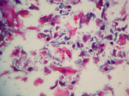
Keratinizing squamous cell carcinoma without necrotic changes (PAP)
Reference Images - Small Cell Lung Cancer
Small Cell Lung Cancer ‐ SCLC (MGG)
Small Cell Lung Cancer – SCLC (MGG). Hypercellular smear with small neoplastic cells witout nucleoli, indistinct cytoplasma and moulding
Reference Images – Granuloma
Aggregates of epitheloid histiocytes forming a granuloma (MGG)
- Content Type